The nucleolus – a known organelle with new tasks
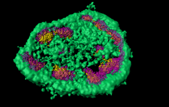
The nucleolus is a well-known cellular structure that is easily visible under a light microscope. This nuclear structure is known as the site of ribosome production. In a recent study, researchers at the Max Planck Institute of Biochemistry in Martinsried, Germany, have shown that the nucleolus is also a site of quality control for proteins. When cells are stressed, proteins tend to misfold and to aggregate. To prevent proteins from clumping, some are temporarily stored in the nucleolus. The special biophysical conditions found in this organelle prevent harmful protein aggregation. The results of this study have now been published in the journal Science.

One would like to believe that the basic cellular processes have already been deciphered and that research can now focus on the details. But even today, new fundamental principles are being discovered through the combination of modern methods. The nucleolus is a nuclear structure that was first described in the 1830s. In the 1960s it was recognized that ribosomes, the protein factories, are produced in this organelle. Researchers have known for some time that protein folding helpers, so-called chaperones, move into the nucleolus under certain circumstances. It has been suggested that this relocation is related to protein production. Now researchers from the Max Planck Institute (MPI) of Biochemistry have shown that the chaperones that move into the nucleolus are bound to stress-sensitive proteins.

As pioneer of chaperone research, F.-Ulrich Hartl and his team have discovered that chaperones are crucial for the correct folding of proteins and play a central role in the quality control of proteins. If protein folding does not work correctly, misfolded proteins can accumulate and clump together. The resulting protein aggregates can often be observed in neurodegenerative diseases such as Alzheimer's, Parkinson's or Huntington's disease.
Mark Hipp, corresponding author of the study and member of F.-Ulrich Hartl's department, comments: “We have been using luciferase as a model protein for many years in order to investigate the mechanisms of protein folding”. Bound to a fluorescent protein, the scientists can see under the microscope whether the protein is correctly folded or if it is misfolded and aggregates. “We were able to show that stressing cells by heating them to 43°C, results in the transport of the misfolded luciferase protein together with the chaperones into the nucleolus.”
The researchers cooperated with the groups of Ralf Jungmann, developer of high-resolution fluorescence methods, and Jürgen Cox, developer of bioinformatic analysis methods, both also located at the MPI of Biochemistry, in order to elucidate the mechanistic details of this process. Together they were able to show that the misfolded luciferase protein behaved differently within the nucleolus than in the rest of the cell. “In the nucleolus, misfolded proteins were kept in a liquid-like state instead of aggregating,” explains Frédéric Frottin, first author of the study. This is possible due to the specific biophysical conditions of this organelle. “Proteins that usually tend to aggregate are stored in a less dangerous form during the stress which protects cells from damage. Once the cell had time to recover, the proteins can be refolded and released from the nucleolus,” continues Frottin. Now, the cells have the capacity to activate further mechanisms that enable the protein to be repaired or degraded. The researchers could also show that this protective mechanism fails if the cell stress lasts too long. “This is a new mechanism that maintains the integrity of the cell,” says Mark Hipp. Maintaining this integrity is ultimately essential to prevent aging and the development of disease.
Original publication:
F. Frottin, F. Schueder, S. Tiwary, R. Gupta, R. Körner, T. Schlichthaerle, J. Cox, R. Jungmann, F.U. Hartl, M.S. Hipp: The nucleolus functions as a phase-separated protein quality control compartment. Science. July 2019
DOI: https://doi.org/10.1126/science.aaw9157
Funding:
The research leading to these results has received funding from the Max Planck Foundation, ERC-grants FP7 GA ERC-2012-SyG_318987-ToPAG and MolMap 680241, as well as the Munich Cluster for System Neurology.


